
Nicholas Skar-Gislinge developed the way we image the nano-world of proteins
Congratualtions Dr. Nicholas Skar-Gislinge!
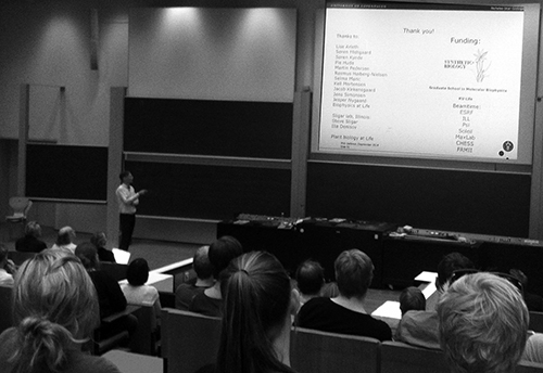
Yesterday, Nicholas Skar-Gislinge defended his PhD entitled: "Developing Nanodiscs as a Tool for Low Resolution Studies of Membrane Proteins". Congratulations!
Painter of Protein Portraits and Silhouettes: With the use of his neutron brush and his X-ray pencil, Nicholas Skar-Gislinge developed the way we image the nano-world of proteins. His work helped to bring other research projects to whole new levels and his imaging tools can prove crucial when designing the sustainable biochemical production machinery of the future.
Academic advisor: Profesor Lise Arleth
Dr. Nicholas Skar-Gislinge
Abstract:
Phospholipid nanodiscs are ~10 nm sized disc shaped particles consisting of roughly 150 phospholipids arranged in a central bilayer, stabilized by two amphipathic protein ”belts” that wrap around the rim of the bilayer. Because they contain a small bilayer leaflet, in which membrane proteins can be incorporated, they can be used as a tool for solution studies of membrane proteins. So far most of the studies of membrane proteins using nanodiscs have been concerning the function of the incorporated membrane protein. However, due to the good control of the size and lipid composition of the nanodisc system, they seem an ideal tool for expanding the use of small angle scattering from studying water soluble proteins, to also include membrane proteins inserted into nanodiscs. This has been the main goal of this thesis.
In order to reach this goal it is necessary to have a detailed understanding of the nanodisc system, without the membrane protein, as well at the self-assembly process in general, and in particular in relation to incorporation of membrane proteins. This was the aim of work done early in this thesis. Here a detailed model for the small angle x-ray and neutron scattering from the empty nanodisc system was derived 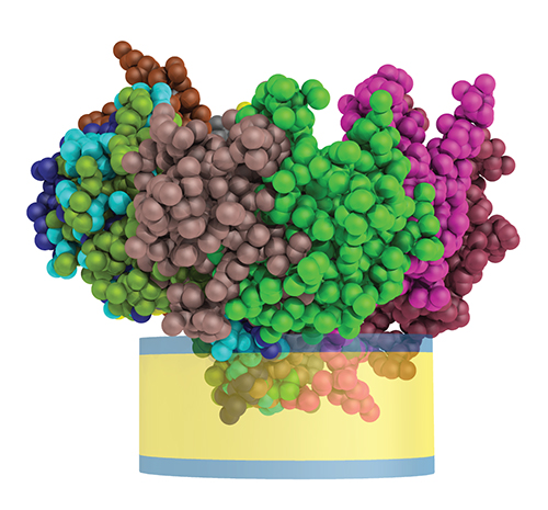 and used to describe the nanodisc system with great detail. The high level of detail was achieved by combining data from both small-angle x-ray and neutron experiments performed on the same samples as well as the incorporation of so-called molecular constraints. Using this model it was found that nanodiscs have an elliptical cross-section, and that the phospholipids in the nanodisc are slightly perturbed, compared to the phospholipids in vesicles.
Later, the self-assembly process of nanodiscs was investigated in more detail by varying the self-assembly conditions. Here it was found that the detergent used for solubilizing the phospholipids has a pronounced effect on the self-assembly process. This may be particularly relevant in order to obtain nanodisc samples of high quality and yield, needed for structural studies using small angle scattering. And which has been a challenge during the whole project.
In the later part of the project the main work has been on obtaining high quality samples as well as conducting SAXS and SANS measurements on these samples, and finally analyzing the measured data. The data analysis was done using a new hybrid approach combining continuous geometric and discrete modeling schemes. The hybrid approach was used to study the placement and orientation of the membrane protein bacteriorhodopsin incorporated into a nanodisc.
Furthermore, the hybrid approach also allows for ab. Initio shape reconstruction of membrane proteins of an unknown shape incorporated into a nanodisc. This approach was investigated for the membrane anchored cytochrome P450 3A4 in the final part of the project
The full thesis can be downloaded from the following link:
https://www.dropbox.com/s/e476q4uv3oupo2l/Thesis_NSG_v2.pdf?dl=0