Glowing DNA invention points towards high speed disease detection
Many diseases, including cancers, leave genetic clues in the body just as criminals leave DNA at the scene of a crime. But tools to detect the DNA-like sickness clues known as miRNAs, tend to be slow and expensive. Now a chemist and a biologist from University of Copenhagen have invented a method that promises to shave days off the lab work done to reveal diseases, using cheap methods and easy to use analytical apparatuses.

Fast, quick and elegant
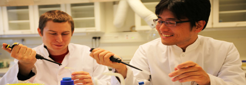
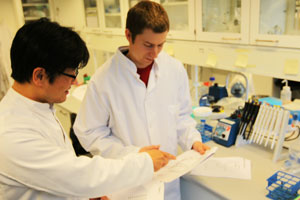
Chemistry researcher Tom Vosch and plant molecular biologist Seong Wook Yang invented a DNA sensor, coupling genetic material to a luminous molecule which goes dark only in the presence of a specific target. Details on their invention, Silver Nano cluster DNA-probes, are published in the high profile scientific journal, ACS NANO and Tom Vosch is understandably proud of the invention.
”We invented a probe that emits light only as long as the sample is clean. That is an unusually elegant and easy way to screen for a particular genetic target”, says Vosch of the Department of Chemistry's Nano Science Centre.
DNA-clues help detecting disease
You could say that the inventors took their cue from crime detection. In murder cases police technicians use DNA to identify the killer. Similarly Individuals with disease are likely to have a unique miRNA profile. Any disease that is attacking a patient leaves this genetic clue all over the victim. And because the profiles of miRNAs vary by type of cancer, finding it proves beyond a reasonable doubt what made the patient sick.
 Gene magnets stick to
opposites
The new detection method
exploits a natural quality of
genetic material. A single DNA
strand is made up of molecules,
so called bases, ordered in a
unique combination. When two
strands join to form their
famous double helix, they do so
by sticking to complementary
copies of themselves.
Likewise, strands tailored to match particular miRNAs will stick to the real thing with uncanny precision. But detecting this union of the strands was only made possible when Vosch and Yang paired their skills.
A real kill switch
Tom Vosch is specialized in studying molecules that light up. Seong Wook Yang is specialized in miRNA. Together they figured out how to attach the light emitting molecules to DNA sensors for miRNA detection. Vosch and Yang discovered, that when these luminous DNA-strands stick with microRNA-strands, their light is snuffed out, giving a very visible indication that the target miRNA is present in the sample. In other words: When the light goes out, the killer is in the house.
Likely to lead to high speed cancer diagnostics
Vosch and Yang tested their Silver Nano Cluster DNA probes with eight different types of genetic material and found that they work outright with six of them. But more importantly, they figured out how to fix the ones, that didn’t. This indicates that their method will work in the detection of almost all types of miRNAs, also in all likelihood for cancer related miRNAs. The most widespread current miRNA detection method requires some 48 hours of lab work from raw samples. The new method can do the same job of detection within a maximum of 6 hours.
Human by accident
A new research method that’s fast, precise and cheap sounds destined for public health-related researches. But the method wasn’t initially meant to find diseases-related miRNAs in humans, says Yang.
”When we started working on the probe, I just wanted to develop a fast and cheap method for detecting plant miRNAs,” explains Seong Wook Yang, and Vosch continues.
”For years I had been making and studying luminous Silver Nano Clusters formed in DNA. Coupling that with Yang’s intimate knowledge of the inner workings of miRNA and the rest of his biological toolboxes turned out extremely fruitful,”concludes Vosch.
Read the Berlingske article based on this report here (in Danish)
Read a summary of the publication resulting from this research here
